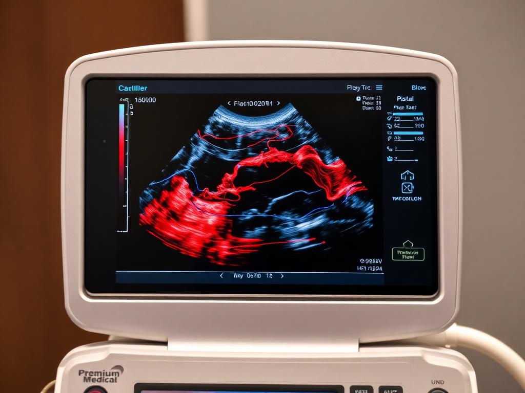
Precise Evaluation of Blood Circulation and Vessel Health
Colour Doppler ultrasound is a specialized diagnostic imaging modality that visually tracks the speed, direction, and resistance of blood flowing through major arteries and veins in real time.
By converting frequency shifts in reflected sound waves into vivid color overlays, Doppler imaging allows clinicians to detect narrowing of blood vessels (stenosis), dangerous blood clots (thrombi), venous valvular insufficiency (varicose veins), and compromised fetal circulation during high-risk pregnancies.
At NOVA Imaging & Diagnostics in Satna, Doppler studies are performed on high-end ultrasound systems and interpreted by Dr. Vikas Garg (MD Radio Diagnosis). We ensure precise hemodynamic waveforms, accurate resistive index (RI) and pulsatility index (PI) calculations, and prompt clinical reports.
When is it Recommended?
Common diagnostic indications
- Obstetric high-risk pregnancy monitoring: maternal hypertension, pre-eclampsia, intrauterine growth restriction (IUGR), or oligohydramnios
- Leg pain, swelling, warmth, or sudden redness to rule out Deep Vein Thrombosis (DVT)
- Visible swollen, twisted veins in the legs (varicose veins) or non-healing leg ulcers (Venous Incompetence study)
- Transient ischemic attacks (TIA), dizzy spells, stroke symptoms, or carotid artery plaque screening (Carotid Doppler)
- Cold extremities, leg cramps when walking (intermittent claudication), or peripheral arterial disease (PAD)
- Refractory high blood pressure or unexplained renal impairment (Renal Artery Doppler)
- Scrotal pain, testicular swelling, or varicocele evaluation in male infertility
Patient Preparation
What you need to know before your test
Obstetric & Peripheral Limb Doppler (Arms & Legs)
- •No fasting is required for limb vascular or pregnancy Doppler scans.
- •Wear loose, comfortable clothing that allows easy access to the arms, thighs, or calf areas.
- •Stay relaxed to ensure steady, representative resting heart rate and blood flow readings.
Renal Artery & Abdominal Doppler
- •Fasting of 8 to 10 hours is strictly required to minimize overlying bowel gas over the deep abdominal aorta and renal arteries.
- •Avoid gas-forming food (beans, milk products, carbonated sodas) the night before the scan.
Carotid & Neck Doppler
- •No dietary restrictions or fasting needed.
- •Avoid wearing tight collared shirts or necklaces around the neck.
Colour Doppler Services Offered at NOVA
All scans and lab investigations are conducted under strict quality protocols with clinical precision in Satna.
Obstetric & Fetal-Maternal Doppler
Evaluation of uterine arteries, umbilical artery, and fetal middle cerebral artery (MCA) blood flow to ensure adequate placental oxygenation and fetal nutrition.
Key Highlights:
Lower Limb Venous Doppler (DVT & Varicose Veins)
Deep and superficial venous mapping to rule out acute blood clots (DVT) and assess saphenofemoral junction competence in varicose veins.
Key Highlights:
Lower & Upper Limb Arterial Doppler
Waveform analysis and luminal narrowing evaluation for peripheral arterial disease, diabetic vasculopathy, and arterial occlusion.
Key Highlights:
Carotid & Vertebral Doppler
High-resolution screening of common, internal, and external carotid arteries in the neck to measure intima-media thickness (IMT) and stroke risk.
Key Highlights:
Renal Artery Doppler
Assessment of main renal arteries, intra-renal resistive indices, and velocity ratios for renovascular hypertension and renal parenchymal disease.
Key Highlights:
Scrotal & Testicular Doppler
Emergency evaluation for testicular torsion (blood flow absence) vs. epididymo-orchitis, as well as pampiniform plexus varicocele grading.
Key Highlights:
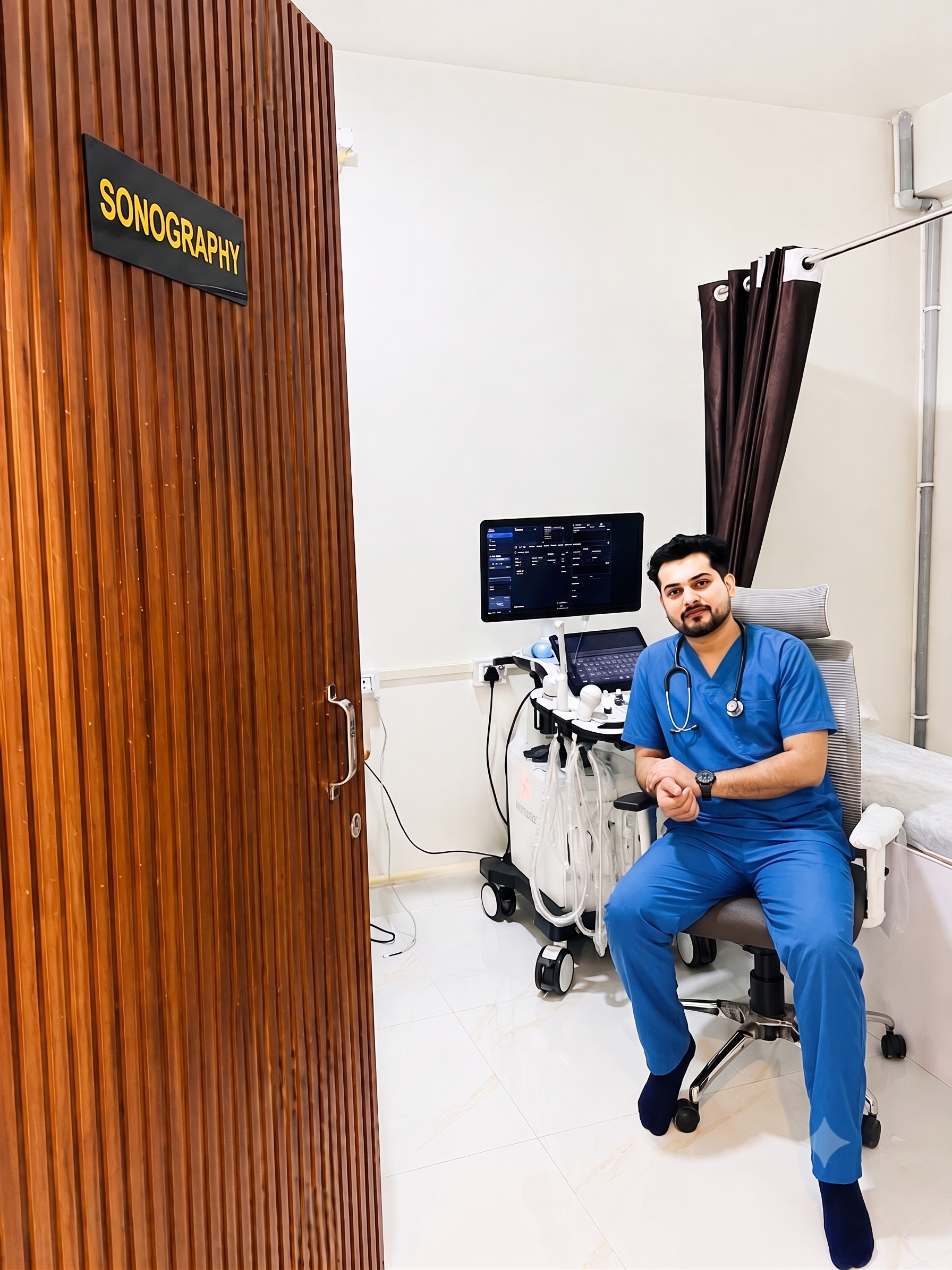
Expert Diagnostic Reporting by Dr. Vikas Garg
MD Radio Diagnosis — MP Medical Council Reg. No. 17920
Every ultrasound scan, colour Doppler study, and digital X-ray at NOVA Imaging & Diagnostics is personally reviewed and reported by Dr. Vikas Garg. We ensure strict adherence to clinical standards, precise organ evaluation, and clear patient guidance in Satna, MP.
Questions About Colour Doppler in Satna
What is the difference between normal ultrasound and colour Doppler?
A standard ultrasound creates grayscale anatomical images of organs, while colour Doppler specifically detects and visually colors blood flow velocity and direction inside blood vessels, helping diagnose blockages, clots, and vascular resistance.
Is colour Doppler safe for the baby during pregnancy?
Yes. Obstetric Doppler is performed adhering to international ALARA (As Low As Reasonably Achievable) thermal and mechanical acoustic indices under Dr. Vikas Garg's experienced supervision.
How is a DVT (Deep Vein Thrombosis) Doppler scan performed?
The radiologist applies ultrasound gel and moves a high-frequency probe along the veins in your leg from the groin to the ankle, gently compressing veins to verify that blood is flowing freely without clots.
How quickly do I get Doppler results?
Comprehensive Doppler reports with detailed velocity waveforms and radiologist findings are delivered on the same day at our Satna centre and shared digitally via WhatsApp/email.
Book Your Appointment
Three quick steps. Our team confirms your slot on WhatsApp or call.
- 1
Patient Details
- 2
Select Services
- 3
Date & Time
NOVA Imaging & Diagnostics, Satna
Diagnostic Services in Satna, MP
Ultrasound / Sonography
2D, 3D/4D, pregnancy anomaly scans, TVS & follicular studies.
Digital X-Ray
Low-dose digital radiography for chest, bones, spine and joints.
Pathology Lab Tests
CBC, LFT, KFT, lipid, thyroid, blood sugar & home sample collection.
ECG (Electrocardiogram)
12-lead digital electrocardiogram for cardiac rhythm & heart health.
Health Checkup Packages
Comprehensive preventive health packages for men, women & seniors.