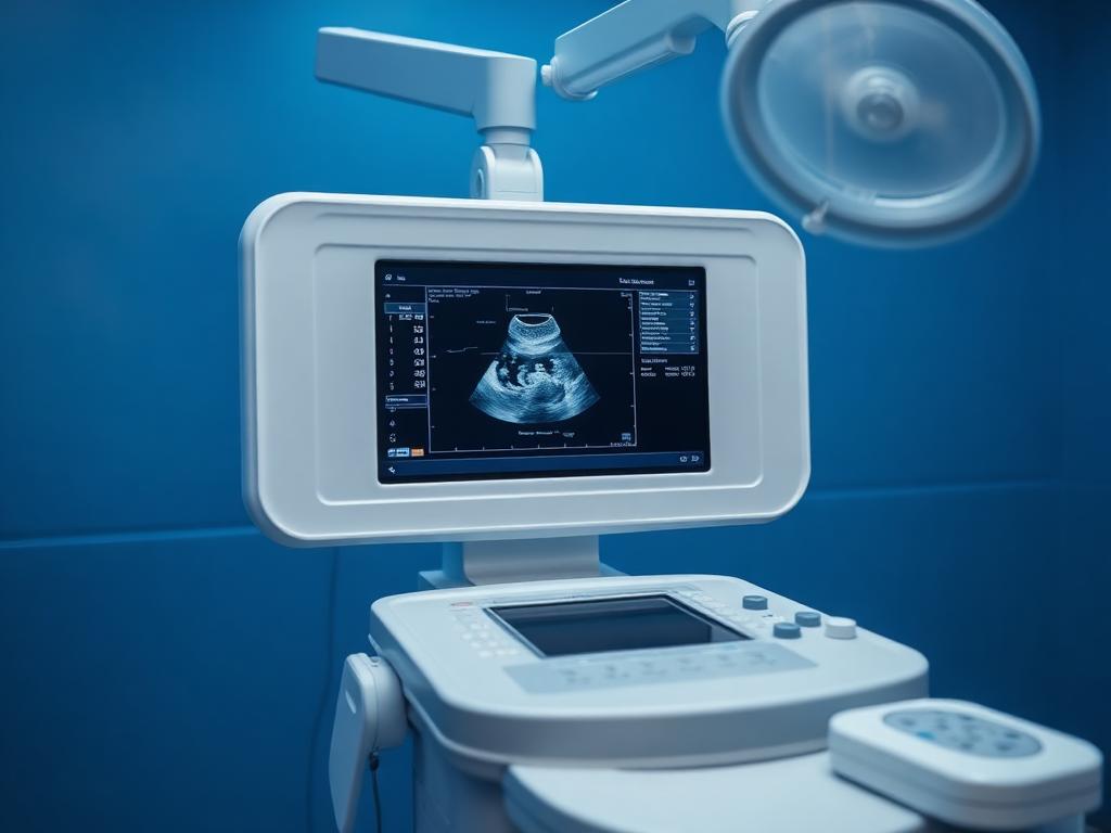
High-Resolution Ultrasound Imaging for Accurate Clinical Diagnosis
Ultrasound (sonography) is a safe, non-invasive imaging technique that uses high-frequency sound waves to generate real-time, detailed images of internal organs, soft tissues, and blood vessels without harmful ionizing radiation.
At NOVA Imaging & Diagnostics in Satna, ultrasound scans are powered by the cutting-edge Mindray Resona i9 Elite ultrasound platform equipped with ZST+ sound speed technology. Every scan is personally performed, reviewed, and interpreted by Dr. Vikas Garg, MD Radio Diagnosis (MP Medical Council Reg. No. 17920).
Whether you require a routine abdominal checkup, comprehensive pregnancy monitoring (NT-NB scan, Level-2 Target Anomaly Scan, 3D/4D fetal imaging), transvaginal sonography (TVS), or small-parts imaging (thyroid, breast, scrotum), we provide clear visual reporting and same-day digital delivery.
When is it Recommended?
Common diagnostic indications
- Abdominal pain, bloating, digestive discomfort, or suspected gallstones and kidney stones
- Obstetric pregnancy monitoring: early dating, Level-1 NT-NB scan, Level-2 Target TIFFA scan, and fetal growth evaluation
- Gynecological issues: pelvic pain, irregular menstrual cycles, fibroids, ovarian cysts, or infertility follicular monitoring
- Urinary symptoms: flank pain, hematuria (blood in urine), urinary tract infections, or prostate enlargement in men
- Swelling, lumps, or pain in the neck (thyroid), breast, scrotum, or soft tissue structures
- Musculoskeletal joint pain, tendon tears, or localized soft tissue inflammation
Patient Preparation
What you need to know before your test
Whole Abdomen & Upper Abdomen Scans
- •Fasting of 6 to 8 hours is required before the scan.
- •Avoid eating solid food, dairy, or fizzy drinks prior to the test to prevent bowel gas obscuring organ visualization.
- •You may sip small amounts of plain water if thirsty.
Pelvic, KUB & Lower Abdomen Scans
- •A comfortably full urinary bladder is essential.
- •Drink 3 to 4 glasses (approx. 1 litre) of plain water 1 hour before the scheduled time and hold your urine.
- •Do not empty your bladder immediately before entering the ultrasound room.
Obstetric Pregnancy Scans (2nd & 3rd Trimester)
- •Fasting is NOT required for routine pregnancy scans.
- •Wear loose, comfortable two-piece clothing.
- •Bring your previous ultrasound reports and doctor prescription cards for longitudinal comparative tracking.
Transvaginal Scan (TVS) & Small Parts (Thyroid, Scrotum)
- •No fasting is needed.
- •For TVS, an empty bladder is preferred; the patient will be requested to void right before the examination.
Ultrasound / Sonography Services Offered at NOVA
All scans and lab investigations are conducted under strict quality protocols with clinical precision in Satna.
Whole Abdomen & Pelvis USG
Complete evaluation of liver, gallbladder, pancreas, spleen, kidneys, urinary bladder, and pelvic organs.
Key Highlights:
Obstetric 3D & 4D Pregnancy Scan
Real-time 3D surface rendering and 4D motion visualization of fetal anatomy, facial features, and well-being.
Key Highlights:
Level-2 Target Anomaly Scan (TIFFA)
Detailed anatomical scan performed between 18–22 weeks to evaluate fetal organ development, spine, heart, and limbs.
Key Highlights:
NT-NB Scan (First Trimester)
Nuchal Translucency and Nasal Bone evaluation performed between 11–13.6 weeks for early chromosomal risk assessment.
Key Highlights:
Transvaginal Sonography (TVS)
High-frequency internal probe scan offering magnification of the uterus, endometrium, and ovaries for infertility & early pregnancy.
Key Highlights:
Small Parts & Musculoskeletal USG
Dedicated high-frequency linear imaging of the thyroid gland, neck lymph nodes, breast, scrotum, and superficial joints.
Key Highlights:
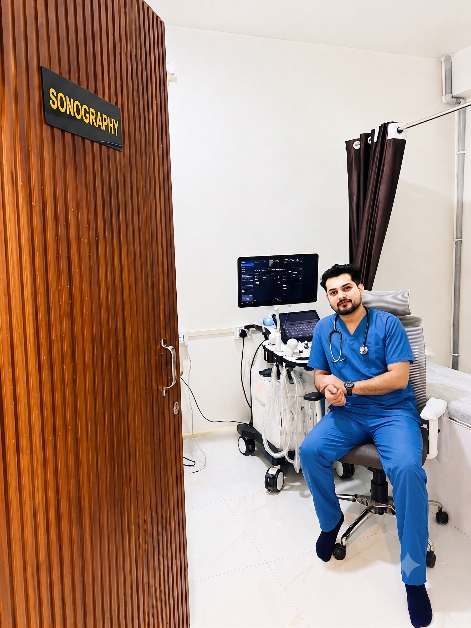
Expert Diagnostic Reporting by Dr. Vikas Garg
MD Radio Diagnosis — MP Medical Council Reg. No. 17920
Every ultrasound scan, colour Doppler study, and digital X-ray at NOVA Imaging & Diagnostics is personally reviewed and reported by Dr. Vikas Garg. We ensure strict adherence to clinical standards, precise organ evaluation, and clear patient guidance in Satna, MP.
Questions About Ultrasound / Sonography in Satna
Is ultrasound safe during pregnancy?
Yes, diagnostic ultrasound is completely safe for both the mother and developing baby. It utilizes sound waves instead of radiation and has been safely used in prenatal care for decades under certified radiologist supervision.
Who performs and reports the ultrasound at NOVA in Satna?
Every ultrasound scan at NOVA Imaging & Diagnostics is personally performed and reported by Dr. Vikas Garg (MD Radio Diagnosis), ensuring experienced clinical judgment, accurate measurement, and clear reporting.
When should I get the Level-2 Anomaly (TIFFA) scan done?
The Level-2 Target Anomaly Scan is ideally performed between 18 and 22 weeks of pregnancy to check the baby's structural development, organs, spine, and brain anatomy in detail.
How long does an ultrasound scan take?
Routine abdomen or pelvic scans take approximately 10–15 minutes. Detailed pregnancy scans such as Level-2 anomaly scans or Doppler studies may take 20–30 minutes depending on fetal position.
How do I receive my ultrasound report?
Physical reports with printed thermal image films are provided on the same day. We also provide secure digital copies via WhatsApp and email for easy storage and sharing with your doctor.
Book Your Appointment
Three quick steps. Our team confirms your slot on WhatsApp or call.
- 1
Patient Details
- 2
Select Services
- 3
Date & Time
NOVA Imaging & Diagnostics, Satna
Diagnostic Services in Satna, MP
Digital X-Ray
Low-dose digital radiography for chest, bones, spine and joints.
Colour Doppler
Vascular, carotid, renal, peripheral & obstetric blood flow studies.
Pathology Lab Tests
CBC, LFT, KFT, lipid, thyroid, blood sugar & home sample collection.
ECG (Electrocardiogram)
12-lead digital electrocardiogram for cardiac rhythm & heart health.
Health Checkup Packages
Comprehensive preventive health packages for men, women & seniors.